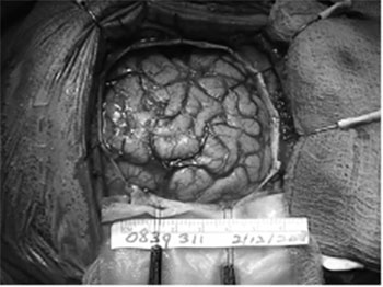
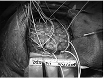
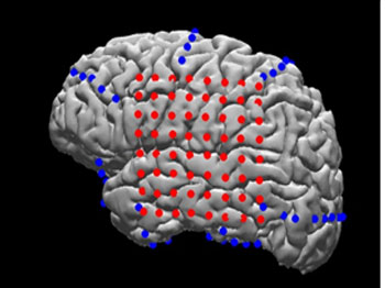
Figure 1. Top: Craniotomy exposing the left temporal and frontal lobes. Middle: Implantation of an 8×8 electrode grid and several surface strips to provide a wide coverage over multiple brain regions. Bottom: MRI reconstruction of the patient’s brain with electrodes overlaid (red: grid array; blue: strip arrays) to allow precise matching of neurophysiological activity to neuroanatomical structures (from Yang et al., Neuroimage, 2012).
Intracranial Electrodes
A number of different methods of intracranial monitoring are possible. Each one has its own set of advantages and limitations. The decision as to which electrode(s) to place within a patient, and which areas of the brain to position the electrodes, are determined on a case-by-case basis that is dependent on the clinical information that needs to be gathered. The most common electrodes that are used for pre-surgical evaluations at the NYU Comprehensive Epilepsy Center are the following:
1) Subdural electrodes
- Subdural electrodes involve the placement of an array of electrodes, known as multicontact grids, directly on the surface of the brain.
- The electrodes are embedded in a clear, flexible material that molds well to the surface of the cortex.
- Local field potentials, which reflect the summated activity from coherent neuronal ensembles, are recorded.
- The main advantage of subdural grids is that they cover a large area of cortex. This includes many areas that are important in cognitive functions, such as language, sensory processing, attention and memory consolidation. Hence, a diverse range of cognitive studies can be performed in patients implanted with these grids.
2) Depth electrodes
- Depth electrodes are placed at precise locations in the brain that are presumed to be potential sites of seizure origin.
- They are often placed symmetrically in the mesial temporal lobes of the brain, including the amygdala and hippocampus. Thus, patients with such arrays are particularly suited to studies investigating working or spatial memory and emotion regulation.
3) Laminar electrodes
- Laminar electrodes enable neuronal activity to be recorded simultaneously in different cortical layers.
- These microarray electrodes allow recordings of local field potentials, multi-unit activity and single-units from 24 contacts spaced 150 microns apart.
- They are particularly valuable for investigating the laminar profile of brain events and creation of current source density profiles to distinguish between feedforward and feedback projections during sensory and cognitive tasks
4) Microelectrode arrays
- The Utah array (Blackrock® Microsystems) contains up to 96 electrodes and enables multichannel, high-density recordings from large populations of neurons.
- Stable neural recordings of action potentials (spikes) and local field potentials can be obtained
- These arrays provide valuable data by providing high spatial resolution within a small area of the brain